 |
|
| node: | IMAGES: What New Coronavirus Looks Like Under The Microscope |
| template: | 4 |
| parent: | Covid-19 / Novel Coronavirus (2019-nCoV) |
| owner: | toxygen |
| viewed by: | |
| created: | 14.02.2020 - 09:13:57 |
| updated: | 14.02.2020 - 09:14:21 |
|
cwbe coordinatez: 101 8333809 8837211 8703087 8710272 ABSOLUT KYBERIA |
| permissions | |
| you: | r, |
| system: | public |
| net: | yes |
total descendants::0
total children::0

COVID-19 coronavirus is seen in yellow, emerging from cells (in blue and pink) cultured in the lab. This image is from a scanning electron microscope.
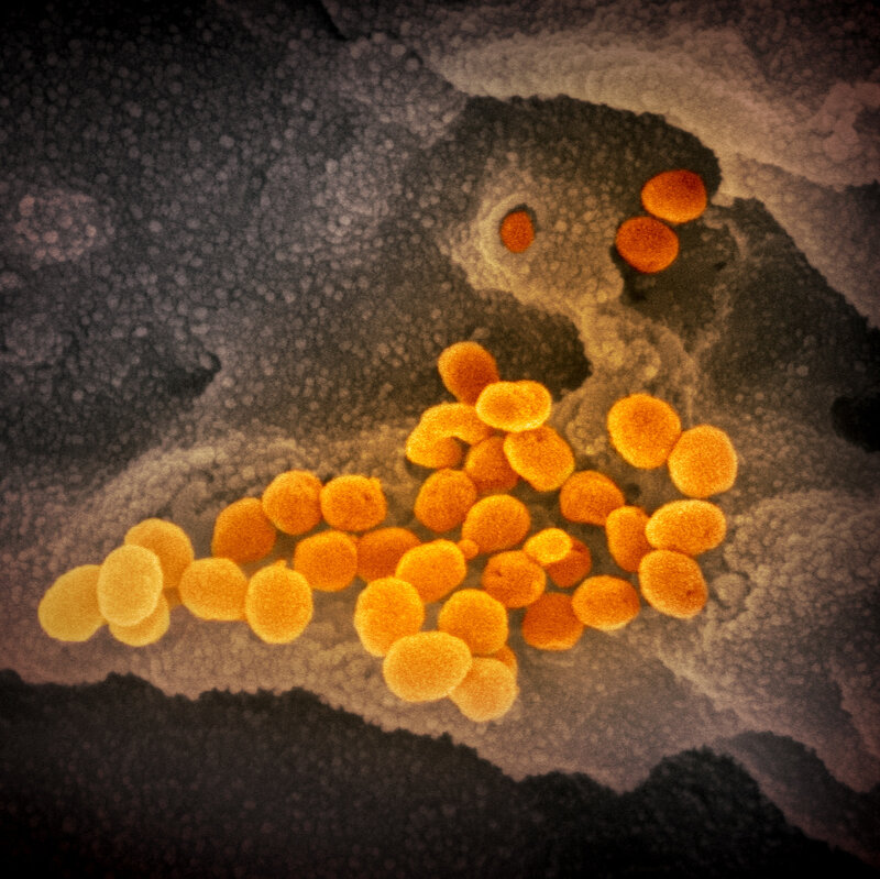
This image from a scanning electron microscope shows, in orange, the coronavirus that causes the disease COVID-19. The virus was isolated from a patient in the U.S. and is seen here emerging from the surface of cells — in gray — cultured in the lab.

In this image from a scanning electron microscope, the new coronavirus is in orange.
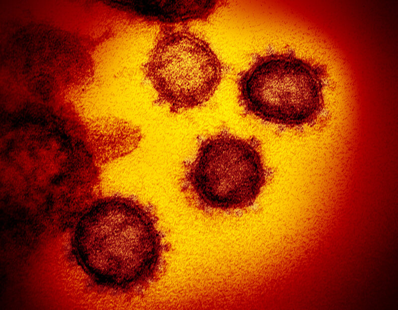
This image of the virus is from a transmission electron microscope.
https://www.npr.org/2020/02/13/805837103/images-what-new-coronavirus-looks-like-under-the-microscope